Interactive OCT and CT image fusion module for clinical otology
Anatomy and pathology
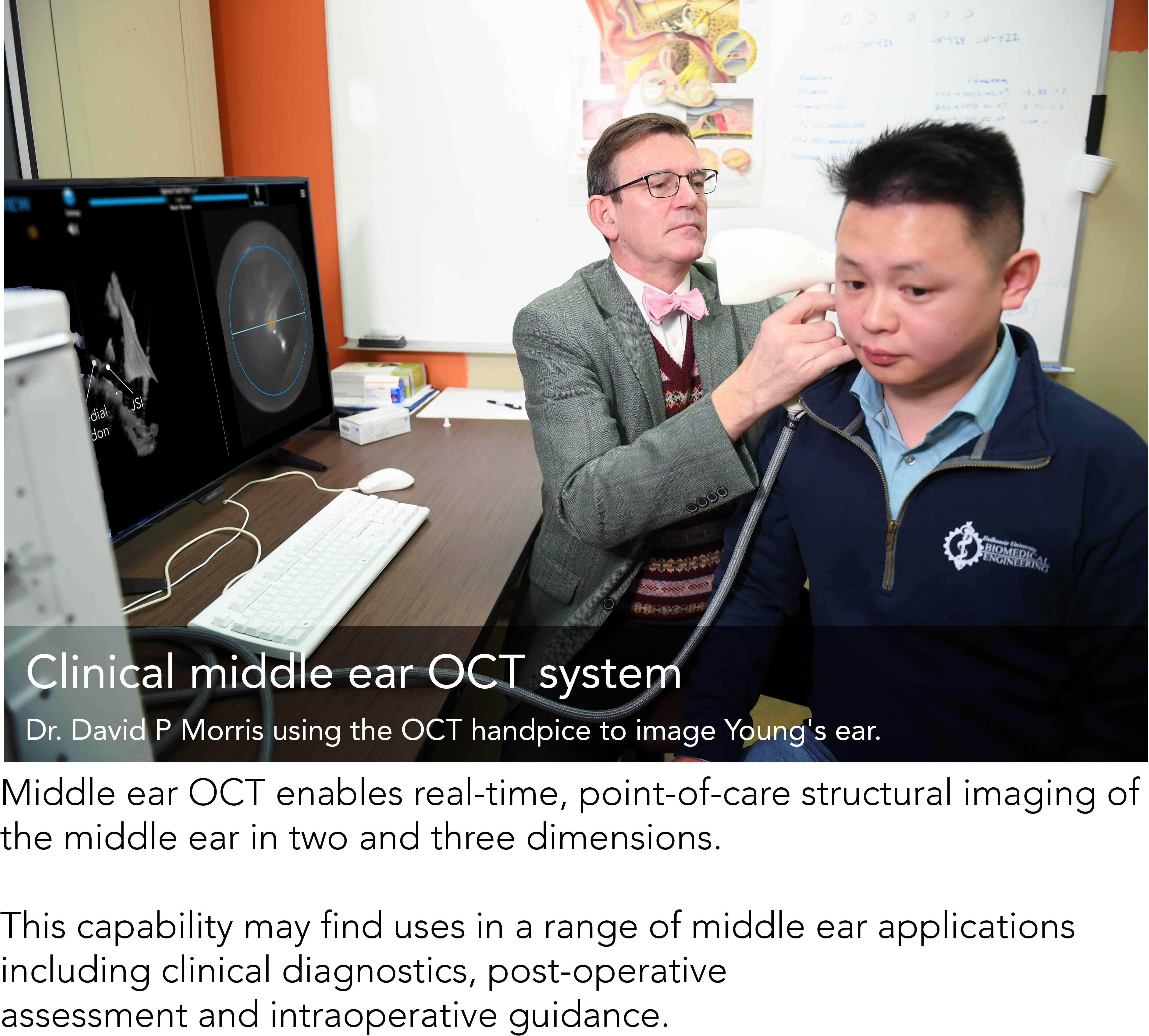
Optical coherence tomography (OCT) is a promising imaging modality for clinical otology. It produces real-time, high-resolution transcanal, transtympanic images of the middle ear (ME) with high soft-tissue contrast. However, its diagnostic applications in otology are limited by its field of view (FOV) and shadowing artifact.
.gif)
This web-based interactive educational module on temporal bone and middle ear anatomy addresses this gap by offering users a platform to familiarize themselves with both the normal anatomy of the middle ear and a selection of pathological cases by the courtesy of Dr. David P Morris and Dr. Nael Shoman, including trauma and cholesteatoma, as observed through in-vivo imaging at our clinic. This module showcases an image fusion technique that augements standard computerized tomography (CT) and OCT for improved clinical interpretation. This results in a composite image that combines the wide FOV and excellent boney delineation of CT with the high resolution and soft-tissue contrast of OCT for better ME images than with either modality alone.
